
Figure 5.
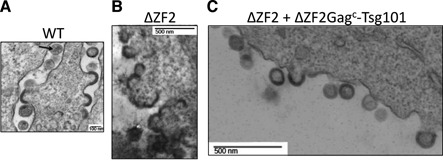
Analysis of the late stages of HIV genesis during trans-complementation experiments. 293T cells expressing pNL4-3 WT (A), pNL4-3 ΔZF2 (B) and ΔZF2 + ΔZF2Gagc-Tsg101 (C) were imaged by electron microscopy (EM). (A) shows WT HIV budding events of immature and mature (arrow) particles. ΔZF2 HIV clearly exhibits budding defects with ‘sticky’ particles with an abnormal morphology (B). The co-expression of ΔZF2 with Tsg101 restores accurate budding with the release of many immature particles (C).