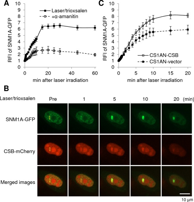
Figure 4.

Response of SNM1A to site-specific DNA ICLs. (A) Quantification of the SNM1A response to laser/trioxsalen, and the effects of α-amanitin on protein recruitment/retention. pSNM1A-GFP was transfected into HeLa cells, which were pre-treated with 6 μM trioxsalen ± 20 μM α-amanitin as indicated, before targeted laser irradiation. The graph shows the RFI of SNM1A-GFP at the microirradiated area relative to unirradiated (background) parts of the nucleus. Each data point is derived from at least 16 independent cells from two independent experiments. Error bars indicate SEM. See Supplementary Figure S5A for representative images. (B) SNM1A and CSB co-localize to sites of DNA ICLs in live human cells. HeLa cells were co-transfected with pSNM1A-GFP and pCSB-mCherry, and co-expressing cells were identified 20–24 h post-transfection. Cells were then microirradiated at the indicated region (yellow box) under the conditions specified. Images of a representative single live cell are shown, along with the merge. (C) CSB coordinates the SNM1A response to DNA ICLs. CSB-deficient (CS1AN-vector) or CSB complemented (CS1AN-CSB) cells were transfected with pSNM1A-GFP and treated with 6 μM trioxsalen for 30 min prior to targeted microirradiation. See panel A for information about the graph, and Supplementary Figure S5B for representative images.
