Figure 2.

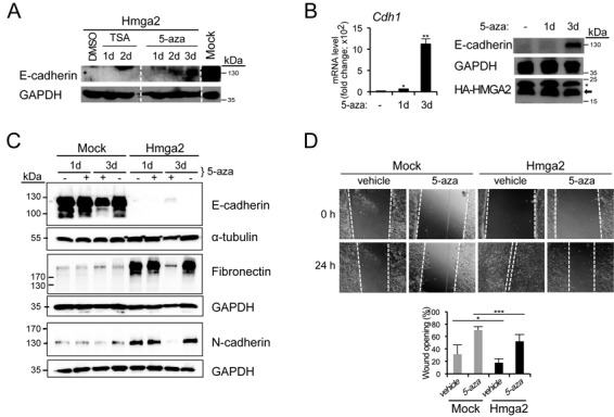
5-Aza-2'-deoxycytidine treatment restores E-cadherin expression. (A) Immunoblots for E-cadherin and GAPDH (loading control) of NM-Hmga2 cells treated with vehicle (DMSO), TSA (100 ng/ml) or 5-aza (20 μM) for 1, 2 or 3 days, and untreated NM-Mock cells. (B) E-cadherin mRNA and protein levels in NM-Hmga2 cells treated with vehicle (−) or 5-aza for 1 or 3 days. Immunoblots for E-cadherin, HMGA2 (arrow) and GAPDH are included. Asterisk indicates unspecific bands. (C) Immunoblots for E-cadherin, fibronectin and N-cadherin of NM-Mock and NM-Hmga2 cells treated with vehicle or 5-aza (20 μM) for 1 or 3 days (+) and untreated cells for the same period of time (−). GAPDH and α-tubulin serve as loading controls. (D) Wound healing assay of NM-Mock and NM-Hmga2 cells treated with vehicle or 5-aza. 5-Aza was added 24 h before the scratch was made (0 h) and measurements were taken 24 h after the wounding. 5-Aza was replenished every day and cells were cultured in the presence of 5-aza or vehicle for total of 3 days. The bar graph shows wound area at 24 h as a percentage of original wound area at 0 h (right panel; mean ± SD from nine fields).
