Figure 5.

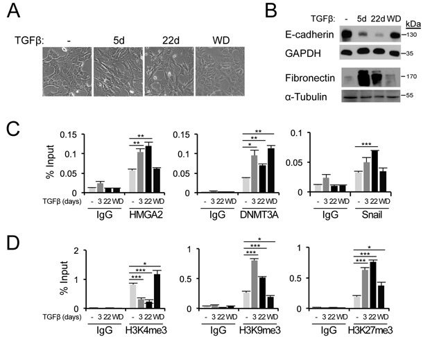
TGFβ-induced EMT is associated with epigenetic changes on the Cdh1 promoter. (A) Morphology of parental NMuMG cells untreated, treated with TGFβ1 (1 ng/ml) for the indicated time periods, or treated with TGFβ1 for 22 days with subsequent withdrawal of TGFβ1 for another 14 days (WD). Scale bar: 50 μm. (B) Immunoblot analyses for E-cadherin and fibronectin protein levels in cells as described in (A). GAPDH and α-tubulin serve as loading control. (C) Binding of endogenous HMGA2, Snail and DNMT3A on the mouse Cdh1 promoter analysed by ChIP-qPCR in cells described in (A). (D) ChIP-qPCR analyses of active H3K4me3 marks and repressive H3K9me3 and H3K27me3 marks at the Cdh1 promoter in cells described in (A).
