
Figure 3.
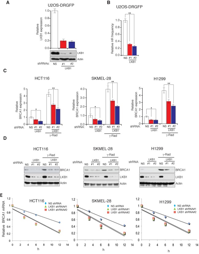
LKB1 regulates BRCA1 mRNA stability. (A) U2OS-DRGFP cell lines stably expressing a non-specific (NS) shRNA or LKB1 shRNAs were analyzed for the expression of LKB1 transcript and protein by RT-qPCR (top) and immunoblot (bottom), respectively. (B) U2OS-DRGFP cell expressing indicated shRNAs was analyzed for HR frequency by monitoring for GFP expression after 48 h of I-Sce1 transfection. Relative HR frequencies under indicated conditions are plotted. (C) The mRNA level of BRCA1 was measured by RT-qPCR in cells expressing LKB1 shRNAs or a non-specific (NS) shRNA that were either unirradiated or gamma-irradiated (20G). Actin expression was analyzed as the internal control. (D) The protein level of BRCA1 was measured by immunoblot analysis in cells expressing LKB1 shRNAs or the NS shRNA that were either unirradiated or gamma-irradiated (20G). Actin was measured as loading control. (E) The half-life of BRCA1 mRNA was measured at indicated time points after irradiation (20 Gray) and treatment with the transcription blocker, actinomycin D (5 μM), in cell lines that either expressed LKB1 shRNA or the non-specific shRNA. The relative BRCA1 mRNA abundance was normalized to actin at the indicated time points and is plotted. The BRCA1 half-lives were significantly different in cells expression NS shRNAs compared to the cells expressing LKB1 shRNAs (P < 0.01). Error bars show standard error mean (SEM). (*P < 0.01; **P < 0.001).