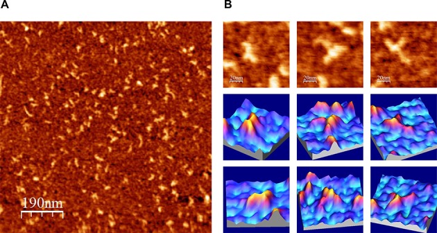
Figure 4.

AFM images of IRES-418 molecules in a folding buffer containing 4 mM Mg2+. (A) Large AFM image (1000 × 1000 nm) of molecules deposited onto a mica-APTES surface. (B) AFM images (100 × 100 nm) of three selected molecules (top row) and their corresponding 3D representations from two view angles (medium and bottom rows). The nominal curvature radius of the AFM tips was 2 nm.
