
Figure 7.
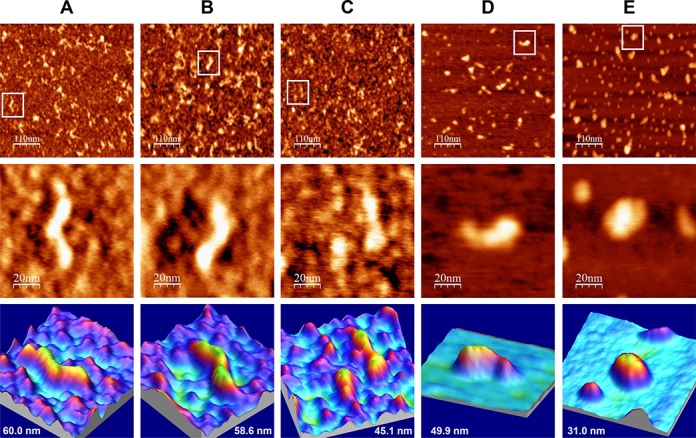
AFM images of IRES-574/miR-122 complexes in a folding buffer supplemented with increasing Mg2+ concentration. IRES-574 RNA molecules were folded in a buffer containing 100 mM Na+, either without Mg2+ (panel A) or supplemented with 2, 4, 6 and 10 mM Mg2+ (panels B, C, D and E, respectively) and incubated with miR-122 (see Materials and Methods). Large AFM images (550 × 550 nm, top row), zoom areas containing a selected molecule (100 × 100 nm images, medium row) and 3D representations of the selected molecule showing its measured length (bottom row) are shown. The medium and bottom row at panel C show an open and a closed IRES-574 conformer coexisting at 4 mM Mg2+, the depicted length corresponding to the open one. Two miR-122 molecules are imaged at panel E (10 mM Mg2+) together with a compact IRES-574 conformer. The nominal curvature radius of the AFM tips was 2 nm.