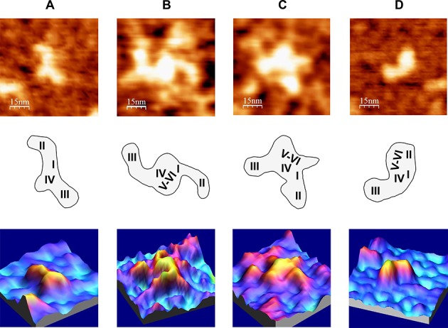
Figure 8.

AFM images of HCV IRES-containing RNA molecules under different ionic conditions, in the absence of miR-122. (A) IRES-418 molecule at 4 mM Mg2+. (B) and (C) Two examples of IRES-574 molecules at 2 mM Mg2+. (D) IRES-574 molecule at 4 mM Mg2+. AFM images (75 × 75 nm) of the selected molecule (top row), schematic representations of the molecule sowing the suggested assignment of IRES domains (medium row) and 3D representations of the selected molecules (bottom row) are shown. The nominal curvature radius of the AFM tips was 2 nm.
