
Figure 2.
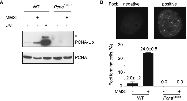
eGFP-Polκ foci formation after MMS treatment. (A) Detection of PCNA-Ub after MMS treatment by immunoblotting. Chromatin-bound proteins were isolated from WT or PcnaK164R cells (MEFs) that were mock treated or continuously exposed to MMS (0.75 mM) for 6 h and separated with SDS-PAGE. After blotting, the membrane was cut below the 38 kDa marker and subsequent antibody staining and ECL was performed separately. The part of the membrane containing proteins with molecular weights >38 kDa (containing PCNA-Ub) was exposed for the maximum time, while the lower part of the membrane (<38 kDa proteins, containing unconjugated PCNA) was exposed only shortly. The asterisk indicates PCNA modified with two ubiquitin moieties. A representative experiment is shown. (B) WT and PcnaK164R MEFs containing eGFP-Polκ were continuously exposed to MMS (0.75 mM) for 6 h after which they were fixed in 4% paraformaldehyde and analyzed with fluorescent microscopy. The average of three independent experiments is shown ±SD.