
Figure 4.
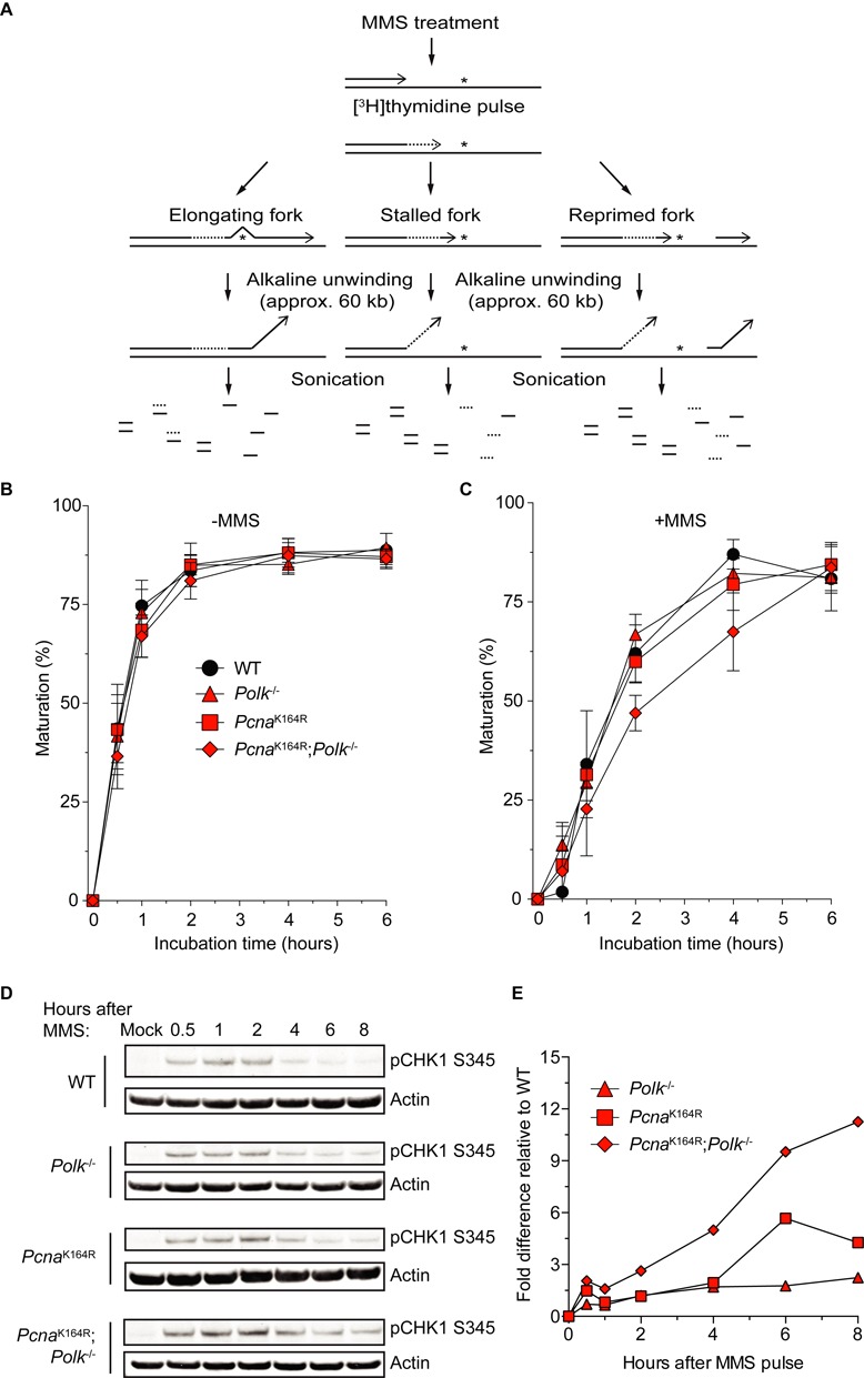
Replication block recovery and ATR/Chk1 activation after MMS treatment of PcnaK164R; Polk−/- MEFs. (A) Scheme of ADU experiment. First, cells are treated with 1.5 mM MMS for 30 min.The asterisks represent MMS-induced replication fork stalling DNA lesions. Then, cells are pulse labeled with [3H]thymidine and chased for different time points as indicated. Subsequently, free DNA ends are locally unwound by alkaline treatment, after which the DNA is sheared by sonication. After binding to hydroxylapatite, ssDNA and dsDNA are eluted separately using appropriate potassium phosphate buffers. Finally, radioactivity is measured in the fractions corresponding to either ssDNA or dsDNA. Adapted from (49). (B) mock-treated MEFs. (C) MMS-treated cells. Average of five independent experiments ±SD. (D) Detection of activated CHK1. p53 knock down immortalized MEFs were treated for 30 min with 5 mM MMS after which they were harvested after indicated time points. Whole cell lysates were analyzed by immunoblotting with a pCHK1S345-specific antibody. (E) Quantification of immunoblot data from (D). All data are relative to WT and normalized to actin levels. Average of two independent experiments.