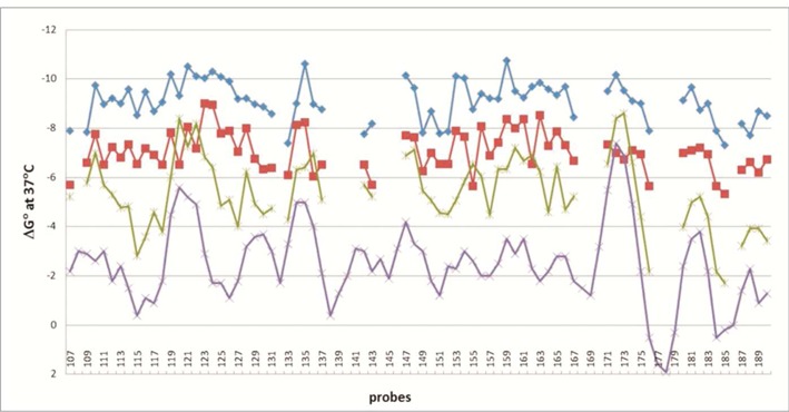
Figure 2.

Comparison of calculated free energies of duplexes formed by DNA (purple line), 2′-O-methylRNA (green line), 2′-O-methylRNA including 2′-O-methyl-2,6-diaminopurine riboside (red line), isoenergetic probes (LNA and 2′-O-methylRNA including 2′-O-methyl-2,6-diaminopurine riboside) (blue line) and complementary single-stranded sequence fragments of RNase P RNA from Bacillus subtilis (RNRspBs). This plot has corrections to the plot on Figure S2 of (62).
