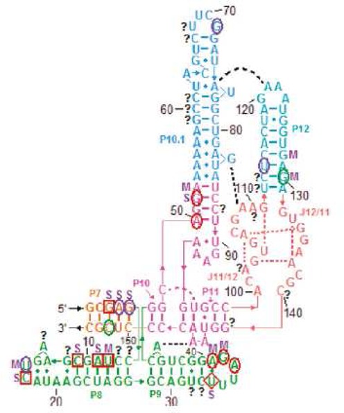
Figure 5.

Binding sites of RNRspBs by isoenergetic probes on microarrays explored in three buffer conditions. Red squares and circles, respectively, indicate strong and medium binding in 140 mM NaCl, 80 mM HEPES, 10 mM MgCl2 and also 135 mM KCl, 25 mM NaCl, 50 mM HEPES, 10 mM MgCl2. Purple circles indicate medium binding only in 135 mM KCl, 25 mM NaCl, 50 mM HEPES, 10 mM MgCl2. The red diamond indicates medium binding in 140 mM NaCl, 80 mM HEPES, 10 mM MgCl2 and strong binding in 135 mM KCl, 25 mM NaCl, 50 mM HEPES, 10 mM MgCl2. Green circles indicate medium binding in 140 mM NaCl, 80 mM HEPES, 10 mM MgCl2. S indicates strong binding in 135 mM KCl, 25 mM NaHEPES, 50 mM HEPES, 1 M NaCl. M indicates medium binding in 135 mM KCl, 25 mM NaHEPES, 50 mM HEPES, 1 M NaCl. Question marks indicate ambiguous binding sites. Reprinted with modifications and permission from (64). Copyright 1999 Macmillan Publishers Ltd.
