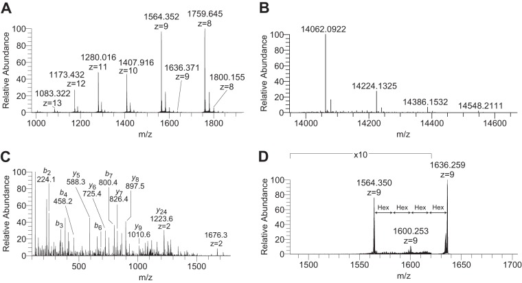
Fig. 3.
Top-down mass spectrometry of the natural Der f 2 allergen. A, Charge-state distribution of Der f 2 following direct infusion and full MS acquisition with 100,000 resolving power at m/z 400. B, Deconvoluted MS1 of multiply charged precursor ions from panel A. Monoisotopic masses of [M+H]+ forms are shown. C, HCD-MS2 of the m/z 1564.35, z = 9 precursor from panel A. The b- and y-type fragments identify the N- and C terminus of Der f 2. All annotated fragment ions are [M+H]+ unless otherwise indicated. D, CID-MS2 of the m/z 1636.37, z = 9 precursors ion from panel A showing the loss of two and ultimately four hexose residues.

