Fig. 1.
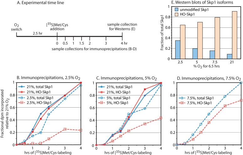
Rate of Skp1 hydroxylation in cells. A, gnt1– cells were transferred to fresh medium and shaken under an atmosphere of the indicated O2 percentage. Metabolic labeling with [35S]Met/Cys commenced at 2.5 h, and cells were harvested at the indicated times and processed for immunoprecipitation using antibodies specific for HO-Skp1 (pAb UOK85) or both HO-Skp1 and Skp1 (pAb UOK77 or mAb 4E1). B–D, time-dependent incorporation of 35S into HO-Skp1 and total Skp1 in cells held at the indicated O2 levels. Data in the same panel are from parallel experiments. Maximal incorporation values into total Skp1 and HO-Skp1 pools after 4 h at 21% O2 were adjusted to 1 to account for variations in antibody pulldown efficiency, and the factor was applied to the parallel low-O2 samples, except for the data in D, which employed the average of factors used for B and C. E, a parallel 4-h sample was subjected to whole cell Western blot analysis using HO-Skp1 (UOK85) and pan-specific (4E1) antibodies to estimate the fraction of Skp1 that was hydroxylated after 6.5 h of exposure to the indicated O2 level. The fraction of Skp1 that was unmodified at steady state in 21% O2 was set at 10% based on the ratio of unmodified to glycosylated Skp1 bands in the wild-type strain Ax3. The unmodified fraction at other O2 levels was derived from the modified fraction via subtraction.

