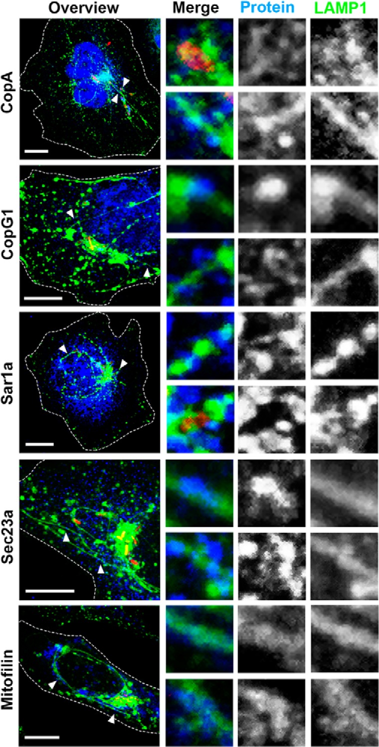
Fig. 7.

Localization of COPI, COPII, and mitochondria in Salmonella-infected cells. HeLa LAMP1-GFP cells were infected with Salmonella WT mCherry (red) and fixed at 8 h p.i. Immunostaining was performed for COPI (CopA, CopG1), COPII (Sar1a, Sec23a), or mitochondria (Mitofilin) (blue), after which cells were imaged using a Leica SP5 CLSM. Images are shown as maximum intensity projections. White arrowheads indicate the structures of interest shown as magnifications for each channel. Scale bar: 10 μm.
