
Fig. 2.
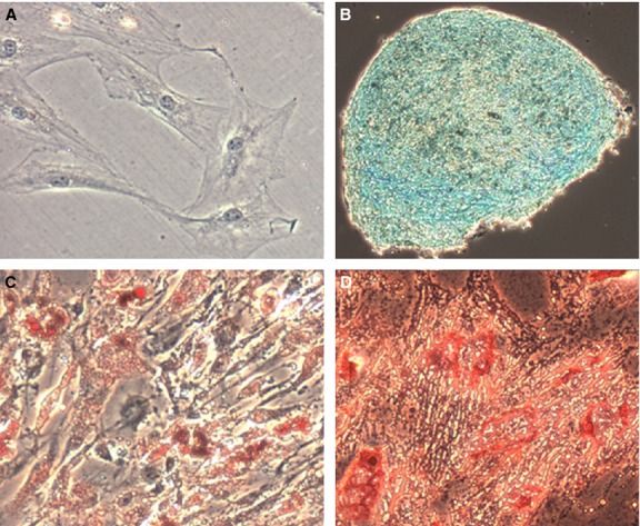
Undifferentated and differentiated ADSCs visualized using microscopy. Original magnification, 10×. (A) Control stain – uADSCs stained with Oil Red O (other controls not shown). (B) Staining with Alcian Blue revealing presence of chondroblasts. (C) Staining with Oil Red O revealing presence of adipocytes. (D) Staining with Alizarin Red S revealing presence of osteoblasts.