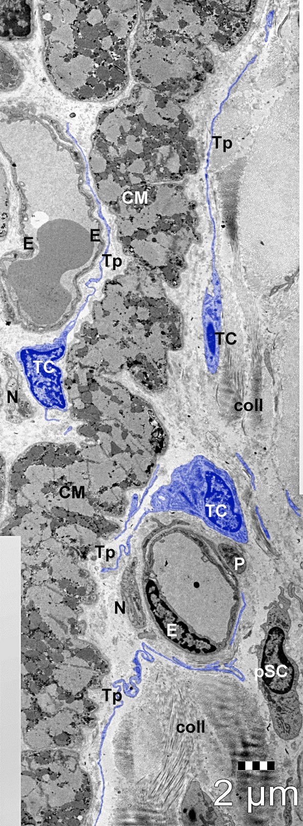
Fig. 2.

General view of human atrial interstitium (8-months-old patient) where telocytes (TC) with telopodes (Tp), endothelial cells (E), pericytes (P), nerve endings (N) and putative stem cells (pSC) could be seen on electron microscopy. Cardiomyocytes (CM); coll – collagen. Bar 2 μm.
