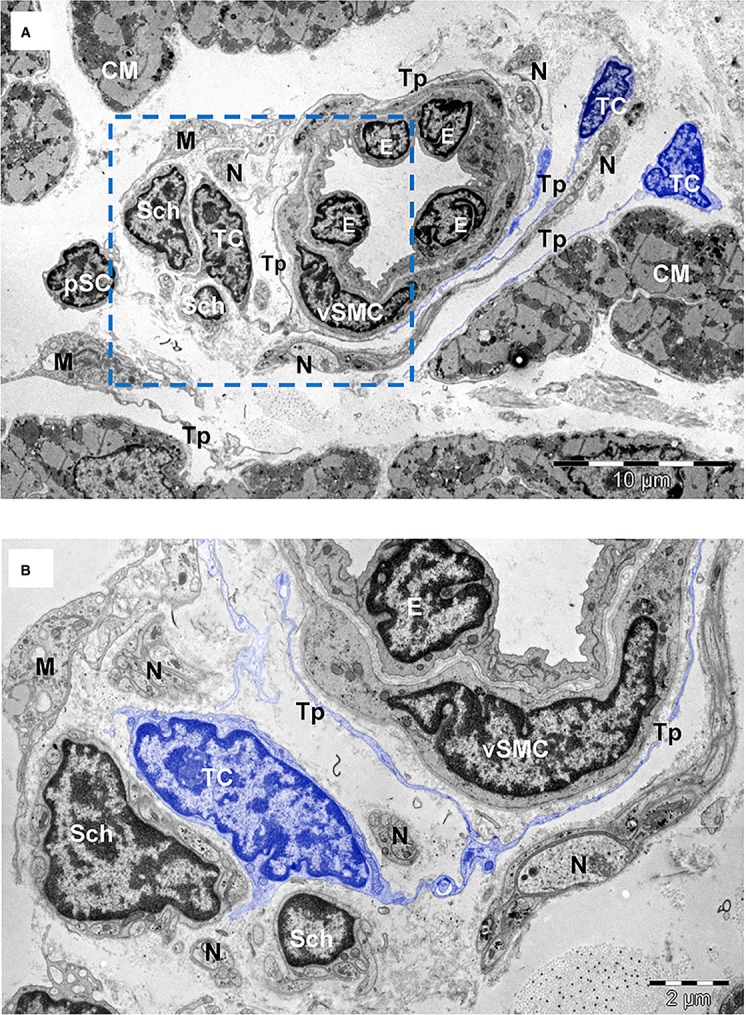
Fig. 4.

(A) TEM image of human atrial interstitium (8-months-old patient) shows telocytes (TC) with long and thin processes (Tp) running around a small artery with endothelial cells (E) and vascular smooth muscle cells (vSMC). There are also visible Schwann cells (Sch), nerve endings (N), putative stem cells (pSC) and macrophages (M). (B) Higher magnification on a consecutive section of the marked area in (A) highlights telopodes (Tp) surrounding the blood vessel in the proximity of Schwann cells (Sch). Cardiomyocytes (CM). Bars 10 μm (A), 2 μm (B).
