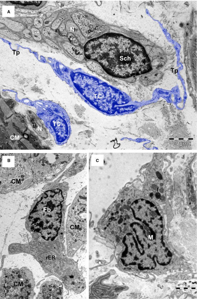
Fig. 6.

TEM images (8-months-old patient) highlight the differences between telocytes (TC) with long telopodes (Tp), and Schwann cell (Sch) (A); the fibroblast (Fb) with abundant rough endoplasmic reticulum (rER) (B) and the macrophage (M) with the cytoplasm filled with lysosomes (l), and coated pits (C). Bars 2 μm (A and B), 1 μm (C).
