Fig. 2.
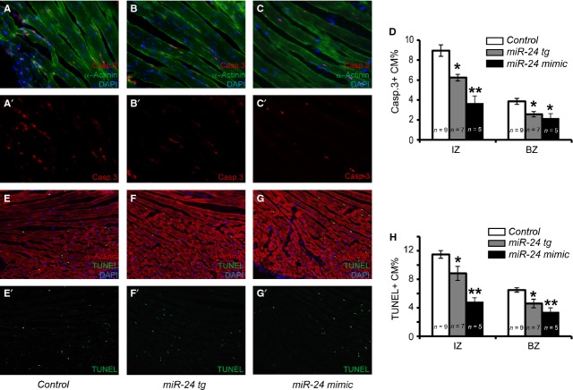
Myh6-miR-24 transgenic mice exhibit reduced cardiomyocyte apoptosis upon myocardial infarction. (A–C') Immunocytochemistry with Caspase 3 (red) and α-Actinin (green) labelling as well as DAPI counterstain (blue) on control (n = 9), miR-24 tg (n = 7) and miR-24 mimic-treated (n = 5) hearts. (D) Quantification of apoptotic cardiomyocytes (Caspase 3- and α-Actinin-positive) in the infarct zone (IZ) and border zone (BZ), with miR-24 tg and mimic-treated hearts showing reduced apoptosis. (E–H) Alternative staining and quantification of apoptotic cardiomyocytes (TUNEL- and α-Actinin-positive) in IZ and BZ, with miR-24 tg and mimic-treated hearts showing reduced apoptosis. *P < 0.05. **P < 0.01.

