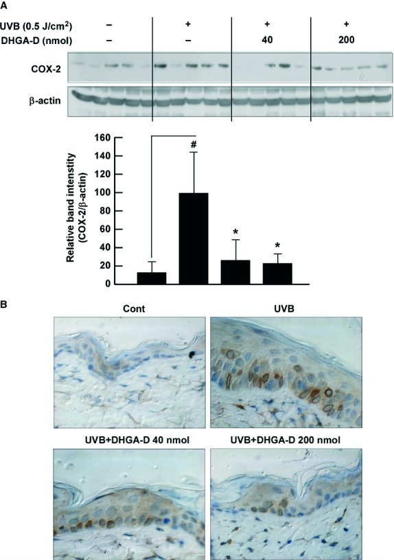
Fig. 6.

Effect of DHGA-D on UVB-induced COX-2 expression in vivo. (A and B) DHGA-D inhibits UVB-induced COX-2 expression in SKH-1 mouse skin. Expression levels of COX-2 and β-actin were determined by Western blot assay with specific antibodies. Each band was densitometrically quantified by image analysis. Results are shown as the means ± SEM (n = 5). The hash symbol (#) indicates a significant difference (P < 0.05) between the control group and the group exposed to UVB alone; asterisks (*) indicate a significant difference (P < 0.05) between groups irradiated with UVB and DHGA-D and the group exposed to UVB alone. In the immunohistochemical analysis, COX-2 is stained brown. Representative photographs of overall immunohistochemical staining patterns from each group are shown.
