
Fig. 4.
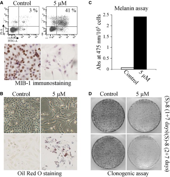
(S)-8 activates multiple pathways in melanoma A375 cells. (A, top) A375 cells were seeded in 6-well plates (105 cell/well) and allowed to attach overnight. The next day cultures were added without/with 5 μM (S)-8 for 48 hrs and then detached and incubated with Annexin-V-Fluos in a HEPES buffer containing PI for 15 min.; the number of apoptotic cells were measured by flow cytometry (FACScan equipment). (A, bottom) Companion cultures were also immunostained with MIB-1 to determine variations of cell proliferation in treated versus untreated cells. (B, top) Phase contrast pictures (magnification ×200) of cultures treated as above showed that (S)-8 caused significant changes in cell density and morphology. (B, bottom) Microscopic visualization of the effects of (S)-8 on accumulation of neutral lipid droplets in A375 cells after fixation and staining with a solution of Oil-Red-Oil (ORO) (magnification ×200). (C) Total melanin content in A375 melanoma cells were assessed spectrophotometrically following 48 hrs treatment with 5 μM (S)-8 (see Materials and Methods) and expressed as absorbance values at 475 nm/105 cells; each column represents the mean ± SD of three separate determinations. (D) For clonogenic assay A375 cells were seeded in 6-well plates (105 cell/well) and allowed to attach overnight. The day after cultures were pre-treated without/with 5 μM (S)-8 for 24–48 hrs. After detachment and counting with a Bürker chamber, viable cells (3 × 10²) were re-plated into new 100-mm dishes and kept with the drug-free medium for additional 7 days, when monolayers were washed and stained with Giemsa to count the number of colonies.