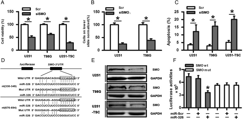
Fig. 2.
SMO knockdown suppressed the biological behaviors of glioma cells, and SMO was a direct target of miR-326. (A) Representative cartogram showing the cell proliferation in glioma cells and tumor stem cells regulated by SMO knockdown. (B) Representative images of in vitro transwell assays of U251 and T98G after transfection with siSMO or scramble RNA. (C) The Annexin V-PI assay reveals increased apoptosis in U251, T98G, and U251-TSC cells following siSMO treatment. The data represent mean ± SE of 3 replicates (*P < .05). (D) Diagram of the seed sequence of miR-326 matched the 3′UTRs of the SMO gene and the design of wild or mutant SMO 3′UTRs containing reporter constructs. (E) Western blot for SMO expression 48 hours after transfection with miR-Scr or miR-326. (F) Luciferase reporter assays in glioma cells after cotransfection of cells with wild-type or mutant 3′UTR SMO and miRNA. The data represent the fold change in the expression (mean ± SE) of 3 replicates.

