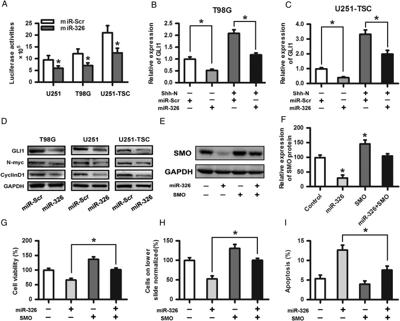
Fig. 4.
Upregulation of miR-326 affected the Hh signaling pathway and rescued the protumor effects of SMO. (A) A plasmid containing the entire SMO coding sequence without its 3′-UTR fragment was transfected into miR-326 or miR-Scr overexpressing cells followed by cotransfection of the reporter containing 8 directly repeated copies of a consensus GLI binding site (8×-GLI) downstream of the luciferase gene to determine the Hh pathway transcriptional activity. The data represent the mean ± SE of 3 replicates (*P < .05). (B) and (C) The overexpression of miR-326 significantly decreased the GLI1 expression, as shown by qRT-PCR with or without N-Shh compared with the miR-Scr-treated group. (D) The Western blot assay indicated that glioma cells transfected with miR-326 efficiently restrained the protein expression of GLI1, N-myc, and CyclinD1 compared with the miR-Scr-treated group. GAPDH was used as a control. (E) and (F) SMO expression levels in U251 cells transfected with SMO and/or miR-326 were assessed by Western blot. Representative cartograms showing the relative expression of the SMO protein between different groups (*P < .05). (G), (H), and (I) Representative cartograms showing that proliferation, invasion and apoptosis are regulated by miR-326 and/or SMO.

