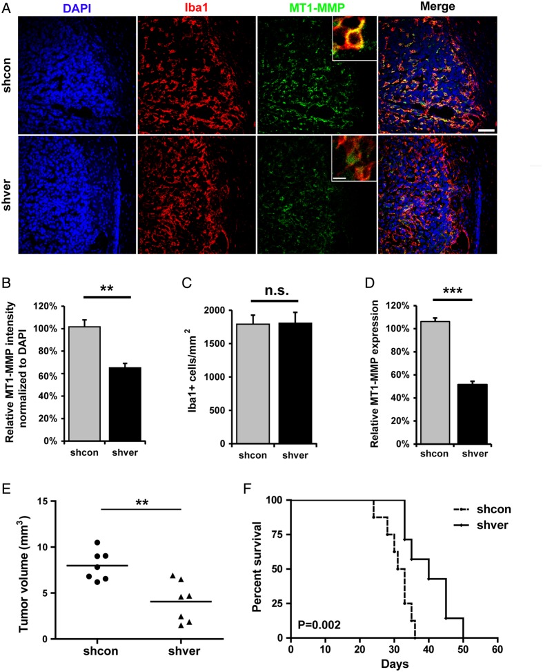
Fig. 3.
Versican-silenced gliomas induced less MT1-MMP expression in microglia, reduced tumor growth, and prolonged survival. (A) Versican knocked down (shver) and control (shcon) GL261 were implanted into wild-type mice, and the glioma tissue sections were stained 14 days after inoculation with DAPI, Iba-1, and MT1-MMP. The image on the right is a merge of all 3 images. Scale bar is 50 μm and 10 μm for the insert. Images were quantified by Image J (n = 6 in each group) to show the difference in MT1-MMP expression normalized to DAPI (B) and Iba-1 positive cell density (C) comparing tissue with shver and shcon GL261 cells. (D) WT mice were injected intracerebrally with shcon or shver glioma cells, and glioma-associated microglia/macrophages were isolated 14 days after glioma cell injection by MACS from both naïve and tumor tissue. Differences in MT1-MMP expression were analyzed by qRT-PCR (n = 3 in each group). (E) shver and shcon GL261 cells were inoculated into WT mice. After 2 weeks, tumor volume was evaluated based on unbiased stereology (n = 7). (F) The Kaplan-Meier curves represent the cumulative survival of mice after shver and shcon GL261 injection (n = 8).

