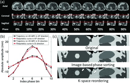
FIG. 4.

(a) Ten-phase k-space reordering 4D-MRI images of the XCAT phantom. Dashed lines are added to assist the visualization of tumor motion. (b) Comparison of tumor motion trajectories between the 4D-MRI and the input signals. (c) Coronal images of the XCAT phantom illustrating the differences between the k-space reordered 4D-MRI and the original XCAT. Background noise is observed in the k-space reordered 4D-MRI (indicated by arrows).
