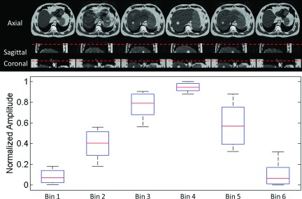
FIG. 7.

Simulated 6-phase 4D-MRI images (top) using the same k-space acquisition scheme and 2D image acquisition mode (interleaves) as in the healthy volunteer study, along with the corresponding motion ranges in amplitude (bottom) for each phase bin.
