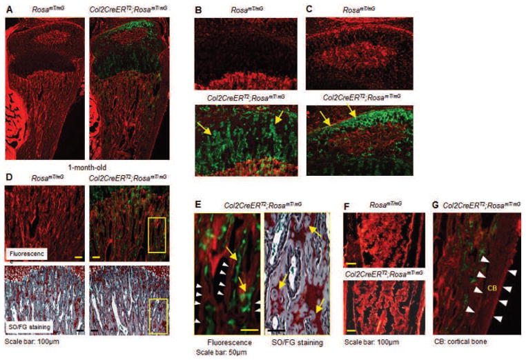
Fig. 1.
Chondrocyte-specific Cre expression in Col2CreERT2 transgenic mice. Col2CreERT2 mice were bred with RosamT/mG reporter mice. The offspring were treated with tamoxifen (TM) at 2 weeks and sacrificed at 4 weeks. Frozen sections were prepared and analysed under fluorescent microscopy. (A–C) A Cre-recombination efficiency over 80 % is achieved in tibial growth plate chondrocytes and articular chondrocytes. The yellow arrows indicate green fluorescent protein (GFP)-positive cells. (D) Some green fluorescent signals are seen embedded in the trabecular bone matrix (upper panel) and Safranin O/Fast green (SO/FG) stained sections show a comparable amount of Safranin O-positive staining inside the trabecular bone matrix (lower panel). In contrast, no green signals are observed on the surface of the trabecular bone. The while arrow heads point to trabecular bone surface and no GFP-positive cells were found on the surface of trabecular bone. (E) Higher magnification pictures of fluorescent and SO/FG staining showed that unremoved chondrocytes are embedded in the bone matrix, suggesting that the GFP-positive cells below the growth plate could be unremoved chondrocytes. However, since the fluorescent picture and SO/FG stained picture are not same section, we could not definitely demonstrate that these GFP-positive cells are indeed unresorbed chondrocytes. Alternatively, these cells may also represent osteoblast precursor cells. The yellow arrows on the left panel indicate GFP-positive cells and the yellow arrows on the right panel indicate unremoved chondrocytes. (F) Only tomato red fluorescence is observed in bone marrow cells of the tibia (far away from the growth plate), indicating that Col2CreERT2 mice do not target osteoblasts. (G) Higher magnificent picture showed that no GFP-positive cells were found in cortical bone (CB) (Cortical bone was marked by white arrow heads). Parts A–C were adapted from Shen et al. (2013) and parts D–F were adapted from Wang et al. (2014).

