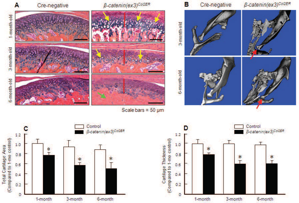
Fig. 2.
Activation of β-catenin induces a TMJ OA-like phenotype. (A) Tamoxifen was administered into 2-week-old β-catenin(ex3)Co12ER mice and Cre-negative control littermates (1 mg/10 g body weight, i.p., daily for 5 d). TMJ samples were harvested when the mice were 1-, 3- or 6-month-old and Alcian blue/H&E staining was performed. Histology results showed increased cartilage degradation in 1-, 3- and 6-month-old β-catenin(ex3)Co12ER mice compared to Cre-negative control mice. Large numbers of hypertrophic chondrocytes were identified in 1- and 3-month-old β-catenin(ex3)Co12ER mice (yellow arrows). Severe subchondral sclerosis was found in 6-month-old β-catenin(ex3)Co12ER mice (green arrow). Reduction in cartilage thickness in 1-, 3- and 6-month-old β-catenin(ex3) co12ER mice is indicated by red bars. (B) µCT images of 3- and 6-month-old β-catenin(ex3)Co12ER mice showed decreased disc space in β-catenin(ex3)Co12ER mice (red arrows) compared to control mice. (C) Total TMJ cartilage area was quantified by tracing the Alcian blue-positive area. Cartilage area decreased 22 %, 41 % and 48 % in 1-, 3- and 6- month-old β-catenin(ex3)Co12ER mice compared to the same aged control mice (*p < 0.05, unpaired Student t-test, n = 5). (D) Total cartilage thickness was quantified by tracing the Alcian blue-positive thickness in the centre of the TMJ cartilage. Total cartilage thickness decreased 21 %, 39 % and 40 % in 1-, 3- and 6-month-old β-catenin(ex3)Co12ER mice compared to the same aged control mice (*p < 0.05, unpaired Student t-test, n = 5).

