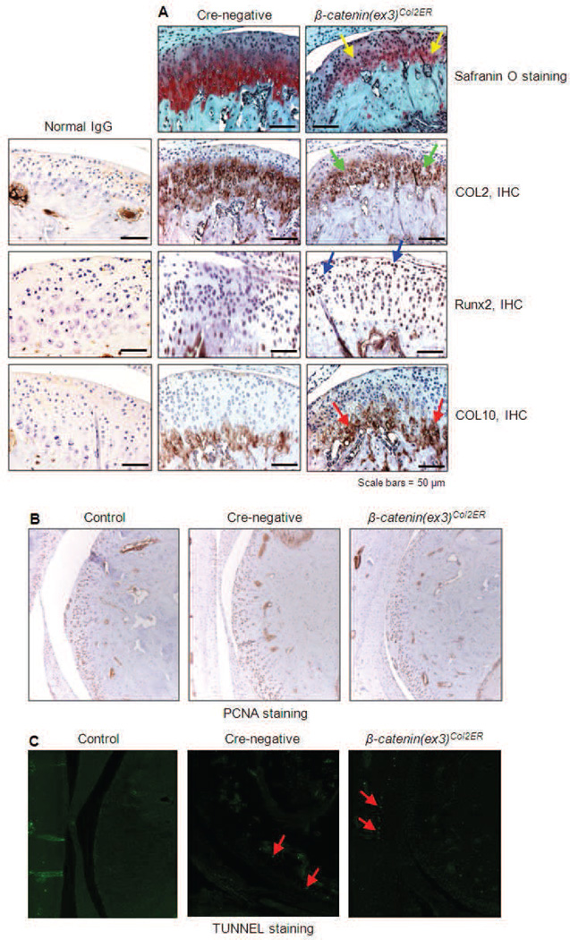
Fig. 3.
Alterations in matrix protein expression, cell proliferation and apoptosis in β-catenin(ex3)Co12ER mice. Tamoxifen was administered when β-catenin(ex3)Co12ER and Crenegative control mice were 2-week-old (1 mg/10 g body weight, i.p., daily for 5 d). TMJ cartilage samples were harvested at 3-month-old. (A) Safranin O/fast green staining was performed to evaluate aggrecan levels. Results showed that aggrecan levels were significantly reduced in β-catenin(ex3)Co12ER mice (yellow arrows). IHC was performed to detect changes in Co12, Runx2 and COL10 protein expression β-catenin(ex3)Co12ER mice. IHC results showed that Co12 expression was significantly decreased (green arrows), and expression of Runx2 (blue arrows) and COL10 (red arrows) was significantly increased in β-catenin(ex3)Co12ER mice. PCNA (B) and TUNNEL (C) staining was performed in TMJ tissues derived from Cre-negative control and β-catenin(ex3)Co12ER mice. Results showed that there were no significant changes in cell proliferation (PCNA staining) and apoptosis (TUNNEL staining) in β-catenin(ex3)Co12ER mice.

