
FIG 5.
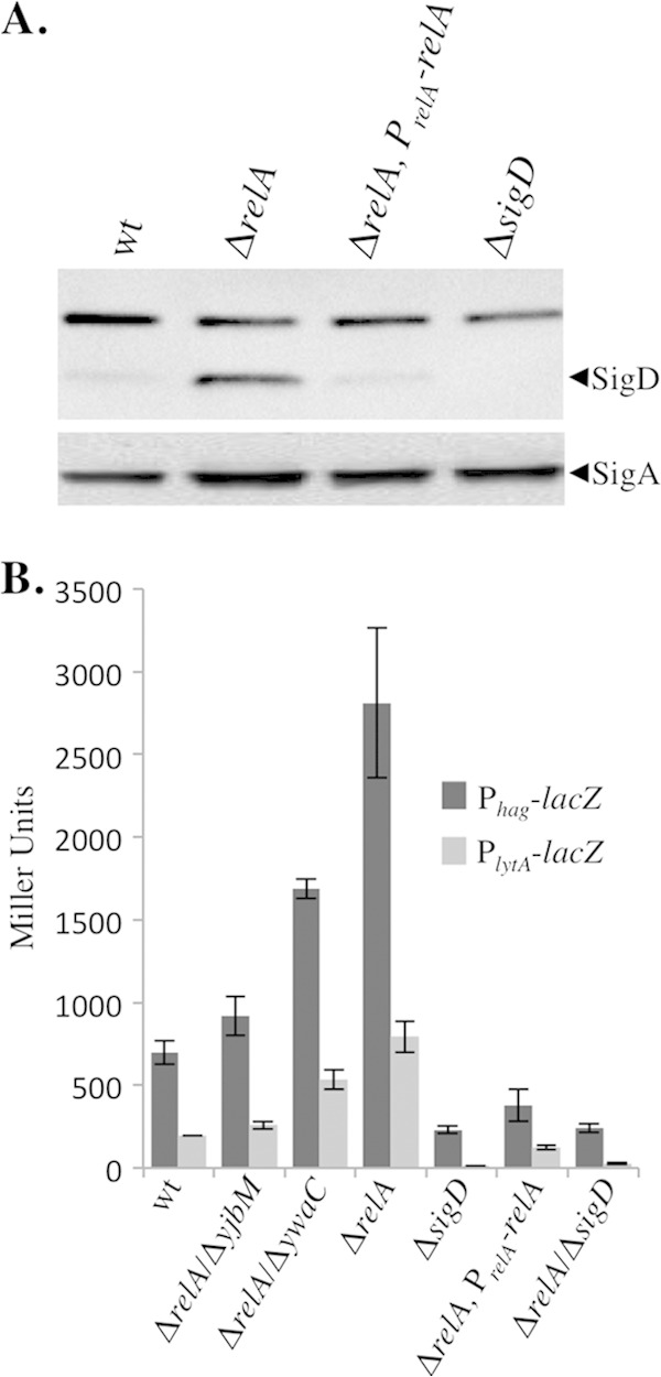
Loss of relA leads to an increase in SigD levels and activity. (A) Comparison of SigD protein levels in wt (BJH001), ΔrelA (BQA009), ΔrelA PrelA-relA (BQA068), and ΔsigD (BQA022) strains. Samples were collected from mid-log-phase cultures grown in CH medium. Proteins from cell lysates were separated by SDS-PAGE and analyzed by Western blot analysis by probing with either anti-SigD or anti-SigA antibody, as indicated. (B) β-Galactosidase assays of Phag-lacZ and PlytA-lacZ transcriptional activities conducted on wt (BJH046 and BJH047), ΔrelA (BQA050 and BQA051), ΔrelA ΔyibM (BQA086 and BQA087), ΔrelA ΔywaC (BQA088 and BQA089), ΔsigD (BQA071 and BQA072), ΔrelA PrelA-relA (BQA073 and BQA074), and ΔrelA ΔsigD (BQA084 and BQA085) strains. Samples were collected from mid-log-phase cultures grown in CH medium. The data shown are the means of three independent replicates with standard deviations.