
FIG 8.
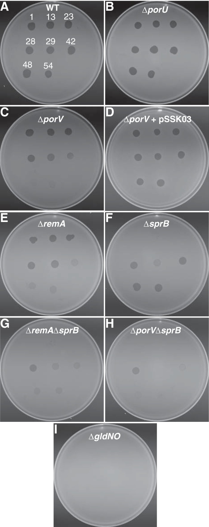
Susceptibility of wild-type and mutant cells to bacteriophages. Bacteriophages (3 μl of lysates containing approximately 109 PFU/ml) were spotted onto lawns of cells in CYE overlay agar. The plates were incubated at 25°C for 24 h to observe lysis. Bacteriophages were spotted in the following order from left to right, as also indicated by the numbers in panel A: top row, ϕCj1, ϕCj13, and ϕCj23; middle row, ϕCj28, ϕCj29, and ϕCj42; bottom row, ϕCj48 and ϕCj54. (A) Wild-type F. johnsoniae CJ1827. (B) CJ2116 (ΔporU). (C) CJ2130 (ΔporV). (D) CJ2130 complemented with pSSK03, which carries porV. (E) CJ1984 (ΔremA). (F) CJ1922 (ΔsprB). (G) CJ1985 (ΔremA ΔsprB). (H) CJ2445 (ΔporV ΔsprB). (I) CJ2090 (ΔgldNO).