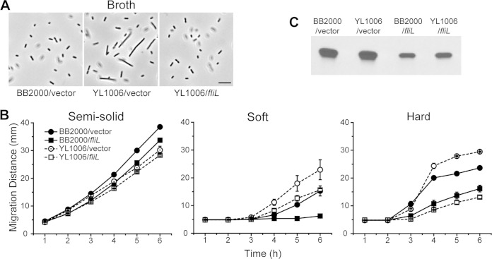
FIG 9.
Cell morphology, migration, and flagellin production of wild-type and ΔfliL mutant cells affected by fliL+. (A) Phase-contrast micrographs of wild-type and ΔfliL cells containing the empty cloning vector and fliL expressed in trans. Scale bar, 10 μm. (B) Migration of wild-type and ΔfliL cells as affected by the agar concentration in the presence and absence of fliL. Shown are the migration distances of the wild-type (solid symbols) and ΔfliL (open symbols) cells containing the empty cloning vector (circles) and fliL expression plasmid (squares) on semisolid, soft, and hard LB agar. Means ± standard deviations from three independent measurements are shown. (C) Comparison of flagellin production in wild-type and ΔfliL cells in the presence and absence of fliL. Shown is an immunoblot to flagellin in wild-type (BB2000) and ΔfliL (YL1006) cells containing empty cloning vector grown on the surfaces of soft LB agar at 37°C for 5 h.

