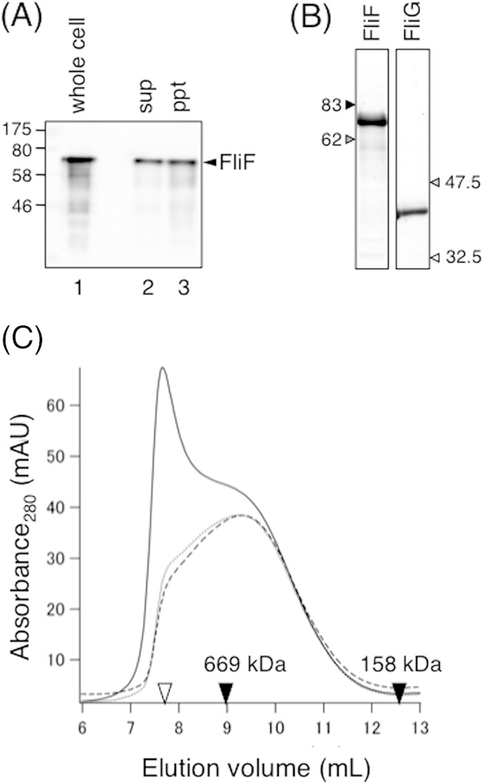
FIG 2.

Size exclusion analysis of purified soluble FliF. (A) Subcellular fractionation of Vibrio FliF overproduced in E. coli BL21(DE3) cells. Cells were disrupted by sonication and then fractionated by ultracentrifugation. The same volume of the supernatant (sup) and pellet (ppt) was analyzed by SDS-PAGE followed by immunoblotting with an anti-FliF antibody. The whole-cell sample was analyzed as a control. (B) Purified FliG and soluble FliF analyzed by SDS-PAGE. The gel was stained with CBB. (C) Elution profiles of purified soluble FliF by analytical size exclusion chromatography using a Superdex 200 column. Purified soluble FliF (1 μM) was subjected to ultracentrifugation, and the supernatant was incubated at 4°C for 24 h. Samples before ultracentrifugation (solid line), immediately after ultracentrifugation (broken line), and after incubation at 4°C overnight (dotted line) were loaded onto the column and analyzed. Open triangle, void volume of the column; closed triangles, elution volumes of the size standards thyroglobulin (669 kDa) and aldolase (158 kDa). mAU, milli-absorbance units.
