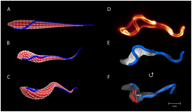
Figure 1. The model trypanosome and a real trypanosome.
(a) Cell body of the model trypanosome without distortion. The elastic network made from vertices connected by springs defines the surface. The blue line connecting a series of vertices represents the flagellum with the helical half-turn. (b, c) Snapshots of the model trypanosome during simulated swimming motion. (d) 3d volume model of a live trypanosome with fluorescently labeled surface. (e) 3d surface model of the cell in (d), with the flagellum highlighted in blue. (f) The same surface model rotated about the horizontal, in order to get a better view on the left-handed half-turn of the flagellum indicated in red.

