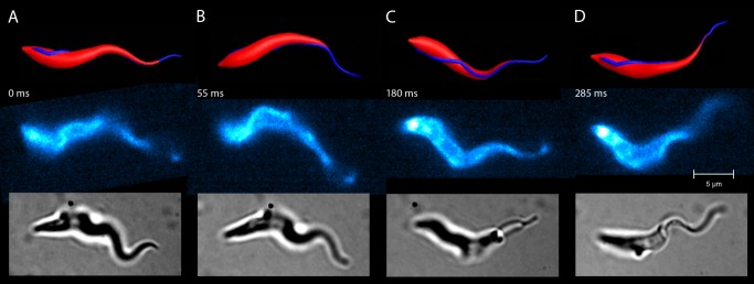
Figure 2. Comparison of the model and a real trypanosome during swimming motion (A–D).

The swimming trajectory and dynamic shape of the simulated model trypanosome (top row) compares well with the forward swimming motion of the real trypanosome (middle and bottom row). Snapshots of the real trypanosome are taken at the indicated times from S4 Video. Fluorescently labelled surface (middle row) or untreated cells (bottom row) were recorded by high speed microscopy (200–500 frames per second). The cells, which exhibit similar speeds and rotational frequencies, show matching cell body conformations at all times over a swimming path of several cell lengths. These periodically repeating shape conformations are initiated by the bending wave passing along the flagellum and determine the trajectory of the swimming parasite.
