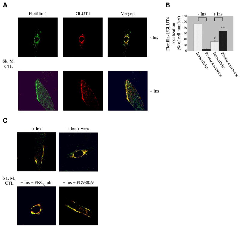
Figure 2.
Flotillin-1 and GLUT4 move to the sarcolemma in response to insulin in a PI3K/PKCζ-dependent fashion in skeletal muscle cells. A) Immunofluorescence. Differentiated myotubes were left untreated or treated with insulin (160 nM for 10 min). Localization of flotillin-1 and GLUT4 was examined using anti-flotillin-1 and anti-GLUT4 IgGs followed by incubation with fluorescent secondary antibodies. B) Quantification. Quantification of localization of flotillin-1 and GLUT4 in skeletal muscle cells before and after stimulation with insulin. Before insulin stimulation, 93 ± 6% of cells expressed flotillin-1/GLUT4 around the nucleus and only 7 ± 1.5% at the sarcolemma. After insulin stimulation, flotillin-1/GLUT4 was found in perinuclear compartments in 32 ± 4% of cells and at the plasma membrane in 68 ± 5% of cells. Values are mean ± SE; n = 100; *P ≤ 0.005 (intracellular localization +Ins vs. −Ins); **P ≤ 0.005 (plasma membrane localization +Ins vs. −Ins). C) Skeletal muscle cells were differentiated for 5 days and treated with insulin (160 nM for 10 min) in the presence or absence of wortmannin (wtm), protein kinase Cζpseudoinhibitor (PKCζ inh.), and PD98059. Expression of flotillin-1 and GLUT4 was detected as in A. Green: flotillin-1; red: GLUT4; yellow: colocalization between flotillin-1 and GLUT4.

