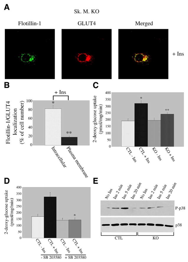
Figure 4.
GLUT4 translocation and glucose uptake are inhibited in caveolin-3 null cells. A) Immunofluorescence. Caveolin-3 null cells were differentiated for five days and treated with 160 nM insulin for 10 min. Cells were then incubated with flotillin-1 and GLUT4 IgGs. Expression of flotillin-1 and GLUT4 was evaluated using fluorescent secondary antibodies. Green: flotillin-1; red: GLUT4; yellow indicates colocalization between flotillin-1 and GLUT4. B) Quantification. Quantification of localization of flotillin-1 and GLUT4 in caveolin-3 null myotubes after insulin stimulation. Flotillin-1/GLUT4 remained localized to perinuclear compartments in 82 ± 7% of cells and moved to sarcolemma in 18 ±3 % of cells after insulin stimulation. Values are mean ± SE; n = 100; *P ≤ 0.005 (intracellular localization KO+Ins vs. CTL+Ins; Fig. 2B); **P ≤0.005 (plasma membrane localization KO+Ins vs. CTL+Ins (Fig. 2B). C) Glucose uptake. Skeletal muscle cells derived from control and caveolin-3 null mice were differentiated for 5 days and treated with or without insulin (160 nM) for 10 min. 3H-2-deoxy-glucose (1 μCi/ml) was added during the last 5 min of incubation. Cells were subsequently solubilized and the 3H content determined by scintillation counting. Values are mean ± SE; n = 9; *P ≤0.005 (CTL+Ins vs. CTL–Ins); **P ≤0.01 (KO+Ins vs. CTL+Ins). D) Glucose uptake. Control skeletal muscle cells were differentiated for 5 days and treated with or without insulin (160 nM for 10 min), in the presence or absence of the p38 MAP kinase inhibitor SB203580 (SB203580 was also added to cells during serum starvation for 4 h). Glucose uptake was determined as described in C. Values are mean ± SE; n = 9; *P ≤0.005 (CTL+Ins+SB vs. CTL+Ins–SB). E) Immunoblot analysis. Differentiated control and caveolin-3 null myotubes were left untreated or treated with insulin (160 nM) for different periods of time (2, 5, and 20 min). Cell lysates were then subjected to immunoblot analysis using an antibody probe specific for the phosphorylated form of p38 MAP kinase. Immunoblot analysis with anti-p38 MAP kinase IgGs (lower panel), which recognize total p38 MAP kinase, indicates that total p38 MAP kinase protein expression was not modified by insulin treatment.

