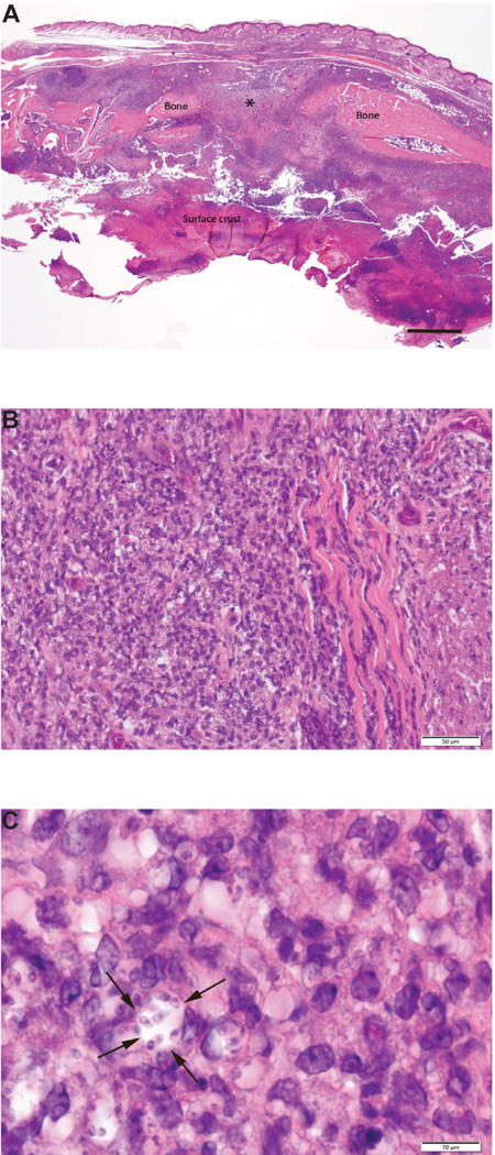
Figure 1.
Photomicrographs of a sagittally sectioned foot of a representative Balb/c mouse sacrificed 25 days PI with L. major and treatment with PBS. Panel A) The footpad epithelium is ulcerated and the surface has a thick crust of cellular debris, inflammatory cells and fibrin. The subcutaneous tissue has severe chronic inflammation. The central area of the metatarsal bone (*) has undergone lysis due to osteomyelitis. H&E, bar=500µm. Panel B) The subcutis has solid sheets of densely packed mixed mononuclear inflammatory cells. H&E, bar=50µm. Panel C) Note that macrophages have Leishmania sp. amastigotes in the cytoplasm. The cell near the center of the photograph has 8 organisms in its cytoplasm (arrows). H&E, bar=10 µm.

