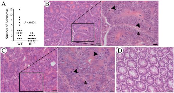
Figure 1. Prothrombin supports colitis-associated adenoma formation.
(A) Quantitation of the total number of adenomas formed per animal after AOM/DSS challenge. Note that mice with a genetically imposed diminution in prothrombin levels to ~50% of normal (fII+/−) developed significantly fewer adenomas. (Horizontal bars represent median values.) Shown are representative H&E stained sections of adenoma tissue harvested from WT mice (B) and fII+/−mice (C). The high-powered insets show the loss of epithelial cell polarity, nuclear pleomorphism, cell piling (*) and frequent mitotic figures (arrowheads) typical of adenomas. (D) A section of unchallenged colonic tissue cut in the same plane is shown for comparison. Size bars represent 20 μm or 10 μm (insets).

