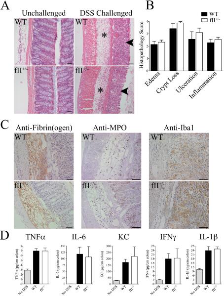
Figure 3. A modest decrease in prothrombin expression does not overtly affect the severity of DSS-induced mucosal damage, inflammatory cell accumulation, or local inflammatory cytokine production.
(A) Shown are representative photomicrographs of unchallenged colonic tissue from WT and fII+/− mice and colonic sections harvested from WT and fII+/− mice following 7 days of DSS challenge. Note that DSS-challenged colons from mice of both genotypes revealed similar degrees of significant mucosal ulcerations (arrows) and inflammatory edema (*). (B) Detailed microscopic analyses using a multiparameter histopathological scoring system confirmed that DSS-challenged colons harvested from WT mice (n = 8) and fII+/− mice (n = 9) showed similar degrees of disease severity for each parameter analyzed. (P values were not significant for each comparison.) (C) Immunohistochemical staining revealed qualitatively similar fibrin(ogen) deposits (brown staining) in both genotyopes that was primarily associated with areas of inflammatory edema. Immunohistochemical staining for neutrophils (α-MPO) and macrophages (α-Iba1) revealed qualitatively similar infiltration with both these cell types in DSS-challenged colons from both genotypes. Size bars in each photomicrograph represent 50 μm. (D) Shown are the levels of inflammatory cytokines measured in colonic homogenates harvested from WT and fII+/− mice immediately following 7 days of DSS challenge (n = 7 per group) as well as levels in colons harvested from unchallenged mice (n = 4). Note that levels of each cytokine measured were significantly elevated in DSS-challenged colons relative to those from unchallenged mice, but prothrombin genotype had no significant effect on local cytokine levels. (P values were not significant for each comparison between DSS-challenged WT and fII+/− cohorts.)

