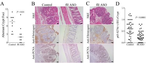
Figure 6. Prothrombin promotes the development of aberrant crypt foci in CAC.
(A) Quantitation of dysplastic foci early AOM/DSS driven CAC performed 1 week after withdrawal of the second DSS course showed that lowering prothrombin levels with a prothrombin ASO to ~50% of normal significantly decreased the number of ACF observed per animal. (B) Shown are representative colonic sections from these same tissues. Dysplastic mucosal lesions consistent with aberrant crypt foci (ACF) were frequently found in mice treated with a control oligonucleotide, whereas the colonic mucosa in fII-specific ASO treated mice was largely healed and appeared normal. Immunostaining revealed significant fibrin(ogen) deposits (brown staining) and PCNA+ colonic epithelial cells (brown staining) in ACF. In contrast, PCNA staining cells were few in number and localized to colonic crypts, and fibrin(ogen) staining was generally scant in more normal appearing mucosa prevalent in fII ASO treated mice. (C) The few ACF that did develop in fII ASO treated mice appeared qualitatively similar to those observed in control mice and exhibited similar degrees of staining for PCNA and fibrin(ogen). (D) Shown is a quantitation of the number of colonic epithelial cell nuclei staining positive for phosphorylated RelA/p65 as a function of the number of intact, properly oriented crypts present within multiple random microscope fields for each mouse. Each data point represents one field and approximately 6 40X fields were assessed per animal. Size bars represent 50 μm.

