
Figure 1. CAA deposition and cerebrovascular dysfunction in young and aged Tg2576 mice.
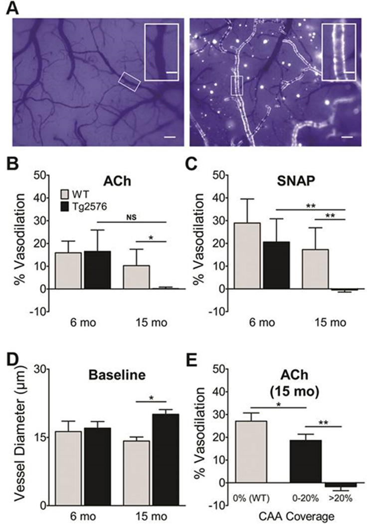
A, CAA deposits in pial arteries of naïve 6 month-old (left) and 15 month-old (right) Tg2576 mice were visualized with the Congo red derivative methoxy-X04 through a closed cranial window. Scale bars: 150 µm (inserts, 50 µm). Vasodilatory responses to ACh (B) and SNAP (C), calculated as percent change in vessel diameter (N=5–7/grp). D, Baseline diameters were similar in young mice; baseline vessel diameter was greater in aged Tg2576 mice. E, Vasodilatory response in aged mice as a function of CAA coverage (N=4 segments/mouse). Data indicate mean ± S.E.M. *: p < 0.05, **: p < 0.01.