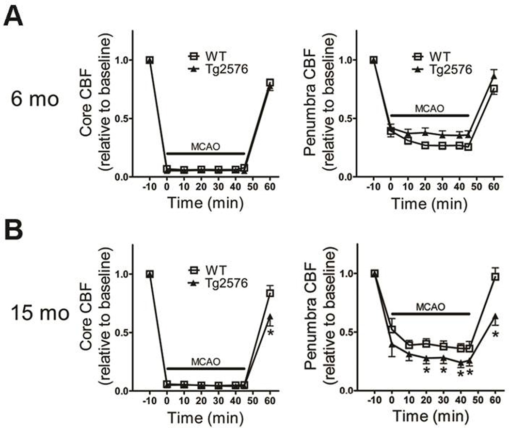
Figure 2. Cerebral blood flow compromise during and after focal cerebral ischemia in aged Tg2576 mice.

Cerebral blood flow (CBF) in the ischemic core (left) and penumbra (right) during focal ischemia was monitored using laser Doppler flowmetery in 6- and 15 month-old mice (N=13–17/grp) (A–B). Data indicate mean ± S.E.M. *: p < 0.05 vs. WT mice.
