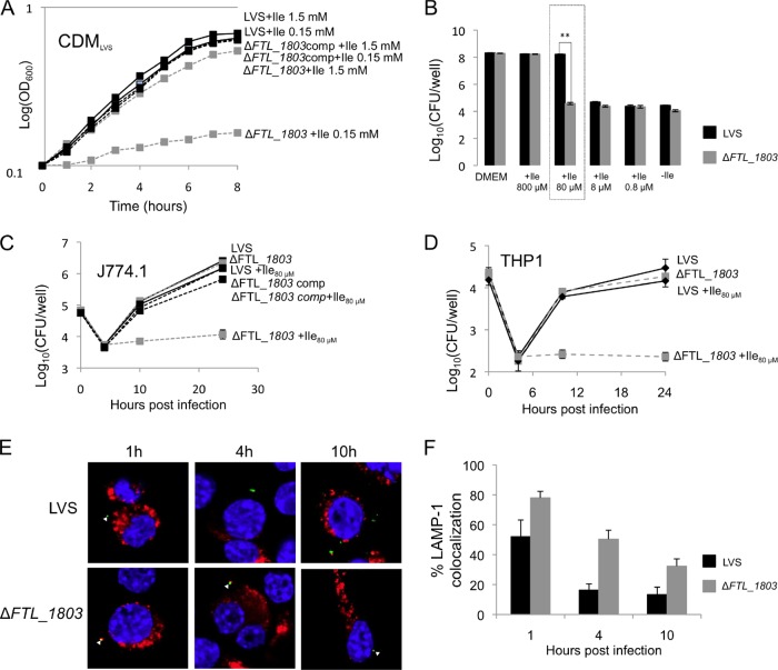
FIG 5.
IleP of LVS (FTL_1803) is required for growth under isoleucine-limiting conditions. (A) Growth in broth. In standard CDM (containing 1.5 mM Ile) (31), growth of the ΔileP mutant (ΔFTL_1803) was slightly affected. When the concentration of Ile was reduced to 0.15 mM, multiplication of the wild-type LVS was essentially not affected, whereas that of the mutant was almost totally abolished. (B, C, and D) Growth in cells. Intracellular multiplication of the ΔileP mutant strain (ΔFTL_1803) was monitored in J774.1 macrophages after 24 h of infection and compared to that of the wild-type LVS in DMEM containing decreasing concentrations of Ile (B). The 80 μM Ile concentration (boxed) was chosen for further analyses. Intracellular multiplication of the ΔileP mutant strain (ΔFTL_1803) was monitored in DMEM supplemented with 80 μM Ile in J774.1 (C) and THP-1 (D) macrophages and compared to that in the wild-type LVS. (E and F) J774.1 cells were incubated for 1 h with the wild-type LVS or the ΔileP mutant strain (ΔFTL_1803), and their colocalization with the phagosomal marker LAMP-1 was observed by confocal microscopy. The phagosomes of J774.1 cells were labeled with anti-LAMP-1 antibody (1/100 final dilution). Cell nuclei were labeled with 4′,6-diamidino-2-phenylindole. Bacteria (white triangles) were labeled with primary mouse monoclonal antibody anti-Francisella (1/500 final dilution). The color images represent wild-type LVS (WT) and ΔileP bacteria (green), phagosomes (red), and nuclei (blue). Quantification of bacteria/phagosome colocalization at 1 h, 4 h, and 10 h for WT and ΔileP strains is shown in the bar graph. **, P < 0.01 (as determined by Student's t test). comp, complemented strain.

