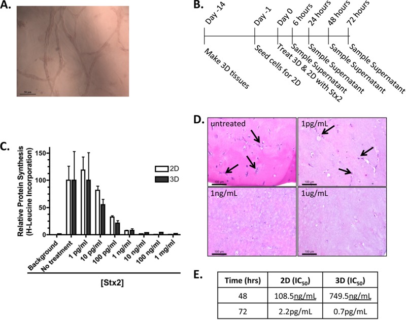
FIG 1.
Shiga toxin-mediated cytotoxicity. (A) Representative image of the interconnecting tubules present in the 3D bioengineered human kidney tissue model. (B) Timeline of tissue formation, treatment, and sampling. (C) A [3H]leucine incorporation assay was used to determine the sensitivity of 2D and 3D cultures to overnight (24- to 26-h) treatment with Stx2 (LPS not removed). (D) H&E image of representative 3D tissues after 72 h of no treatment or treatment with noted concentrations of Stx2. Arrows indicate the hematoxylin-stained cells within the matrix. (E) IC50 values at specific time points for 2D and 3D tissues.

