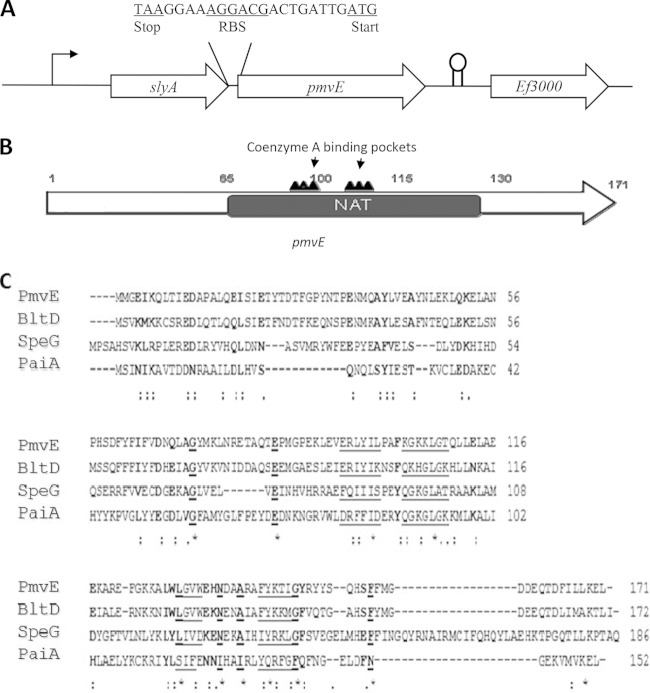
FIG 1.
(A) Genetic organization of the pmvE chromosomal region. Large arrows represent the genes which compose the open reading frame and its genetic environment, and their orientation shows the transcriptional direction. The intergenic sequence between slyA and pmvE is presented. (B) Structure of the pmvE gene showing the N-acetyltransferase (NAT) conserved domain harboring the two highly conserved coenzyme A binding pockets. (C) Alignment of the amino acid sequences of PmvE from E. faecalis, BltD and PaiA from B. subtilis, and SpeG from E. coli (CLUSTALW). Strictly conserved amino acids are underlined and in bold, and conservatively substituted residues are in bold. The underlining indicates residues that are involved in binding CoA.

