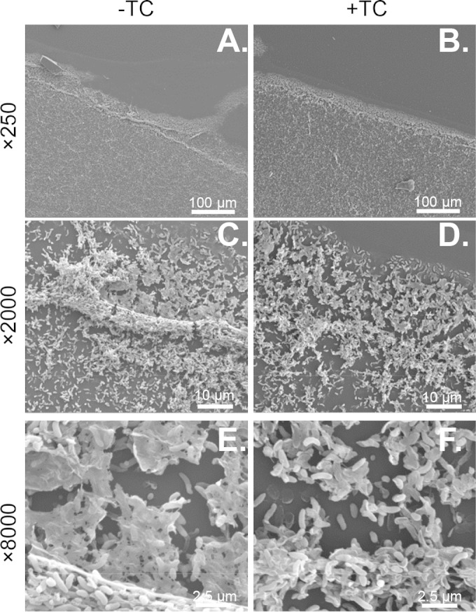
FIG 3.

Representative images of V. cholerae biofilms imaged using scanning electron microscopy after 24 h of growth on a 55- by 55-mm glass coverslip followed by 1 h of exposure to 1 mM TC (B, D, and F) or no TC (A, C, and E). Images are at ×250 (A and B), ×2,000 (C and D), and ×8,000 (E and F) magnification.
