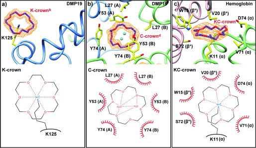
Figure 3.

18-Crown-6 binding modes. The upper part of each panel illustrates the structure of the molecule, the lower part is a representation, where dashed lines represent hydrogen bonds and the red semi-circles are hydrophobic contacts. a) In the K-crown binding mode, a single lysine binds the CR axially. b) Hydrophobic and π-orbital containing side-chains interact laterally with CR. No residues interact with the central region of CR, which commonly but not always coordinates two water molecules. Letters in parenthesis indicate the chain ID. c) In the mixed KC-crown binding mode, the CR is coordinated axially by a lysine, while hydrophobic and π-orbital containing side-chains interact with it laterally. Letters in parenthesis indicate the chain ID, with asterisk indicating symmetry equivalents.
