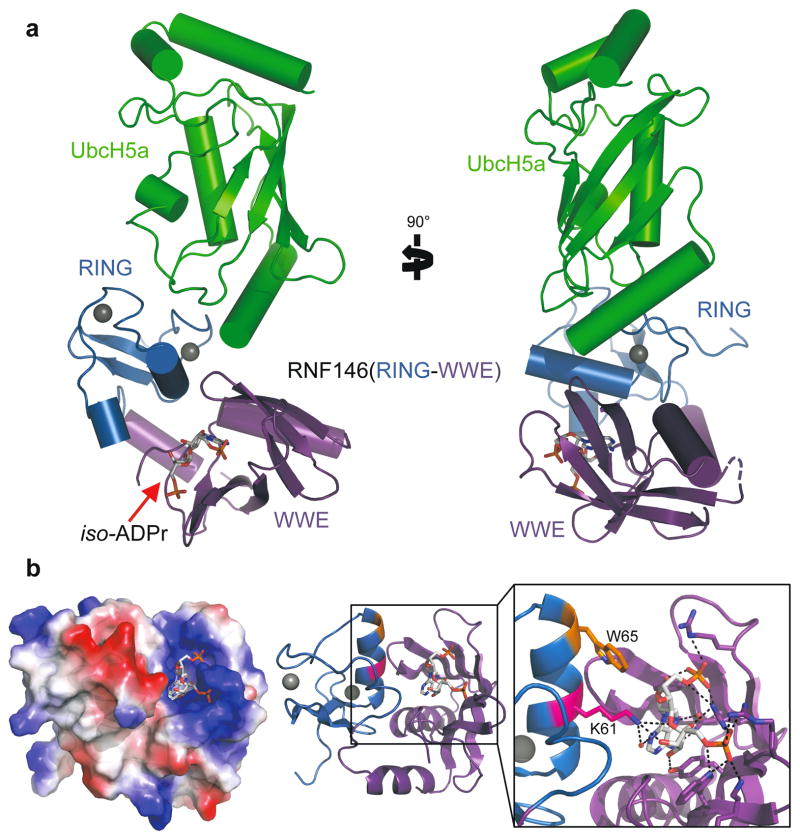
Figure 2. Crystal structure of the RNF146(RING-WWE)/UbcH5a/iso-ADPr complex.
a. Cartoon representation of the RNF146/UbcH5a complex with RING domain colored blue, WWE domain colored purple, and UbcH5a colored green. Zn2+ ions are shown as grey spheres, and the iso-ADPr ligand is represented as sticks. b. The RNF146/iso-ADPr interface. (Left) Surface electrostatic view of RNF146(RING-WWE), showing the iso-ADPr/PAR binding pocket; (Center) same view, cartoon representation; (Right) close-up view of iso-ADPr pocket. Polar contacts between protein and the ligand, iso-ADPr, are indicated by dashed lines; RING residues K61 (magenta) and W65 (orange) are highlighted.

